


Amazing Ways 3D Printing Helps Save Babies’ Lives
Doctors use 3D printed models to practice intense, life-or-death surgeries

A quick note: Thanks for bearing with me during my absence. I had a busy few weeks here — no childcare when my son’s preschool was closed for a break, followed by a family visit, followed by my husband AND son getting covid. It looks like we’ll be back on track, mostly, this week and will be posting weekly again. Fingers crossed. I can only make time to write here when I have childcare and no other pressing priorities.
When people talk about 3D printers, sometimes they think about their gadget-maker at home. Or perhaps they think about a fantastical future where a machine can program a meal, one extruded layer at a time.
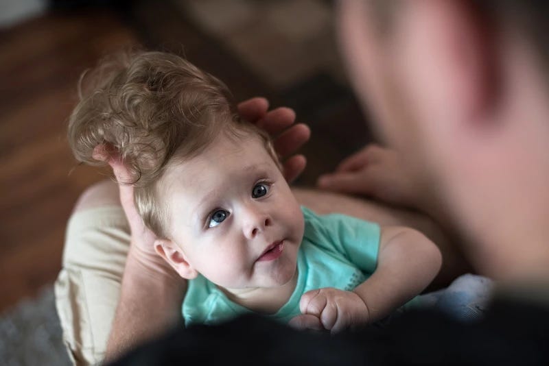
Usually, I think about baby Bentley.
When I was reporting as a freelancer for Vice in 2016, I came across a story of an incredible use of 3D tech in pediatric surgery. It was about how doctors at Boston Children's Hospital had used their new 3D printing program to print models of a 4-month-old’s brain to plan a complex surgery that would determine the course of his life.
You see, Bentley Yoder wasn’t expected to survive birth. His parents found out at 20 gestational weeks that Bentley’s brain hadn’t formed as expected. Part of his brain was outside his skull — a birth defect called encephalocele. The condition is rare, serious, and often fatal during pregnancy or infancy.
It is a vast under-simplification to say the odds were stacked against Bentley. His parents decided to birth him and expected him to die that day. But he took a breath, then another, and 36 hours later he was still alive.
The Washington Post also did a profile on the family and Bentley’s mom Sierra Yoder told them, “We looked at the nurse and we said, 'What do we do now?'” She told the paper that doctors said to go home and arrange hospice care.
Yet four months later — after days that felt like weeks and weeks that felt like years — there was a glimmer of hope. The portion of the brain outside Bentley’s skull that had been expected to die was still live tissue, and not only that — Bentley was using that part of his brain. It needed to be protected, and that meant getting it into the protective cradle of the skull.
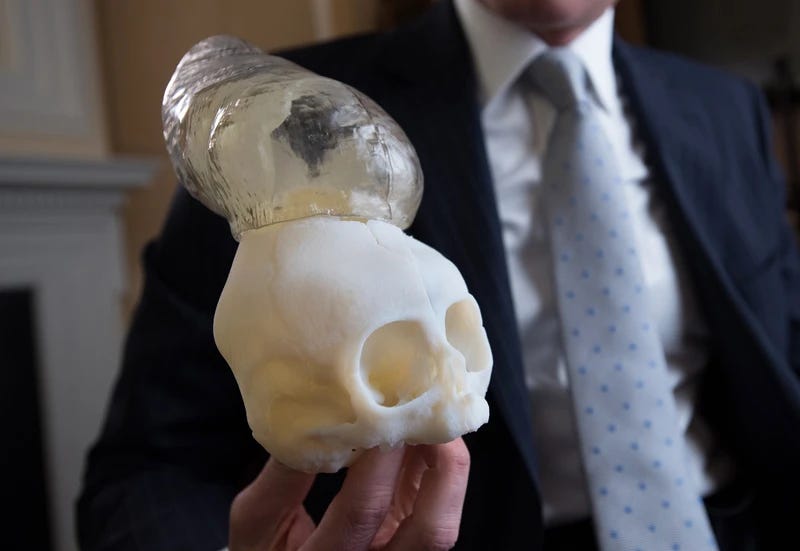
To prepare for this surgery, usually his doctors would use MRIs or CT scans to have a 2D representation of the blood vessels, bone, and brain structures they’d encounter in the surgical theater. But thanks to their hospital’s 3D printing program, they had those scans and also were able to use them to create a physical model of Bentley’s brain.
Seeing a flat image of a brain and being able to hold and turn a patient’s brain is quite a different experience, Dr. John Meara, now plastic surgeon-in-chief at Boston Children’s Hospital, told me in our 2016 interview.
"When you print out this model, it's an amazing opportunity for me to actually look at the anatomy and look at the skull from different angles and actually hold it in my hand and spin it around," he said in our interview. "It's very critical for these kinds of cases. They're rare, and each one is a little different, so you have to make a treatment plan just for these patients."
The surgery was a success and Bentley was “doing fantastic” almost a year afterward, Meara told me.
As of August 2021, Bentley was doing well and managing multiple challenging disabilities, according to his parents’ support Facebook page. I wasn’t able to find any 2022 updates on him online, which honestly is probably good news. He’s a big enough name in the medical community that no news probably means things are stable. I wish the best for him and his family.
Many other hospitals have used 3D printing technology for pediatric, neonatal, and in-utero surgeries. Yes, you read that right — surgeries done on fetuses in their parent’s uterus.
Here’s a 2021 report from 3D printing B2B publication The Additive Report:
“‘The 3D reconstruction of the fetus can really educate the surgeon on the real-life shape, size, and location of the spinal lesion, as well as prepare the surgeon to have the appropriate equipment ready to treat this condition surgically,’ said Samer Elbabaa, M.D. Elbabaa, the medical director of pediatric neurosurgery at Orlando Health, added, ‘It’s a level of detail that we are not able to see in traditional imaging, but that is extremely valuable in these cases where we cannot actually see the defect ahead of surgery.’
To create the models, Orlando Health works with the prototyping and 3D printing team at Digital Anatomy Simulations for Healthcare LLC (DASH), the Orlando, Fla., company that developed the technology.
Crude, single-color items have been 3D-printed in the past, but DASH has taken the process to the next level, developing technology to enhance MRI and ultrasound images taken throughout the pregnancy to reconstruct accurate curves and edges. DASH prints the high-resolution models with multiple colors and materials, allowing surgeons to see details such as skeletal structure, nerve and vascular anatomy, and fluid sacs in the spine and brain caused by spina bifida.”
Amazing, isn’t it?
I wish no one ever needed these surgeries. But I’m thankful this developing technology of 3D printing is helping doctors save babies who desperately need them.
Sources:
https://www.vice.com/en/article/aek49k/hospitals-are-using-3d-printing-to-save-childrens-lives
https://pubmed.ncbi.nlm.nih.gov/28008429/
https://pubmed.ncbi.nlm.nih.gov/27079366/
https://www.facebook.com/BentleyStrong1101/
Excellent article